Benign Paroxysmal Positional Vertigo (BPPV)
Benign Paroxysmal Positional Vertigo (BPPV)
Benign Paroxysmal Positional Vertigo (BPPV) is the most common cause of vertigo cases. BPPV is characterised by dizziness (mild to severe) with movements of the head, often from lying to sitting and rolling over in bed (Wietske, Bruintjes, Oostenbrink & van Leeuwen, 2005).
Near 50-70% of BPPV cases are idiopathic, meaning without a known cause. Other secondary causes can include, a blow to the head, which represents about 7-17% of the cases, viral labryrinthitus (Inner ear inflammation) causes 15% of BPPV cases, Menieres disease (5%), Migraines <5%, and inner ear surgery <1% (Parnes, Agrawal & Atlas, 2003).
Patients who suffer from this condition can also suffer from loss of balance, blurred vision, nausea and vomiting. Around 80% of patients experience rotary vertigo (the sensation of being on a roller coaster) while 47% experience a floating type sensation (Kentala, 2000). These episodes last less than a minute.
This condition is easily diagnosed and easily treatable with a few simple diagnostic and treatment maneuvers. This condition is self-limited and usually resolves by itself within 6 months. Many patients are so anxious they go to great lengths to avoid having a vertigo episode that they are completely unaware that the condition has spontaneously resolved.
BPPV is easily diagnosed and easily treatable with a few simple diagnostic and treatment maneuvers #physio #vertigo Share on X
Benign Paroxysmal Positional Vertigo (BPPV) is the most common cause of vertigo cases #dizzy #physio #vertigo Share on X
The anatomy of the vestibular system
To have a good understanding of vertigo one must understand the anatomy and physiology of the vestibular system. Simply put, the vestibular system detects where the head is in space by monitoring acceleration and deceleration movements of the head (Parnes, Agrawal & Atlas, 2003).
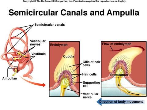
The inner ear contains 3 semicircular canals. These canals are known as the anterior, posterior and horizontal or lateral canal. Each of these canals may be affected, however the posterior canal is the most gravity dependent canal of the vestibular system and therefore is the most commonly affected (Parnes, Agrawal & Atlas, 2003). When a person who has BPPV moves from lying to sitting or vice versa, the accumulation of the calcium carbonate/crystals in the cupula travel into the semicircular cannels (this is called canalithiasis) (Parnes, Agrawal & Atlas, 2003), and by so doing send messages to the brain that the head is moving when it isn’t.
The accumulation of calcium carbonate (small crystals) gathering together in the cupula or the lumen of the semicircular canals (Figure 1), happens benignly or from trauma to the head (Stambolieva & Angov, 2006).
The most common cause of BPPV in the population under the age of 50 years is due to trauma from the head (Herdman, Tusa & Herdman, 2000), however in the aging population the cause is due to degeneration in the vestibular system (Herdman, Tusa & Herdman, 2000).

The semicircular canals of the inner ear.
Posterior canal BPPV
Posterior canal BPPV is the most common type of BPPV due to its gravitationally dependent semicircular canal. Because of the posterior canals orientation this type of BPPV does not resolve easily. This canal hangs more inferiorly and with normal daily head movements it is unlikely the free floating crystals would find their way out of the canal and back into the cupula. In this canal when the free floating crystals move away from the ampulla this causes the crystals to move in an ampullofugal direction (away from the ampulla) which causes an excitatory or stimulatory response. This response induces nystagmus and vertigo (Parnes, Agrawal & Atlas, 2003).
Lateral or horizontal canal BPPV
Lateral or horizontal canal BPPV makes up for about 30% of BPPV cases and resolves much more quickly than posterior canal BPPV (Parnes, Agrawal & Atlas, 2003). Because of the orientation of the lateral canal, lateral BPPV resolves much more quickly as the free floating crystals can easily make their way out of the lateral semicircular canal with normal head movements (Parnes, Agrawal & Atlas, 2003). If a person with lateral BPPV turns their head towards the affected ear this will cause the free floating crystals to flow towards the ampulla which creates an ampullopetal endolymph flow (flow of the fluid in the ear to go towards the ampula). This causes a stimulatory response and then creates a geotropic nystagmus.
Nystagmus means fast pace involuntary eye movements and geotropic means towards the ground. If the patient turns their head to the non-affected ear side the endolymph will flow in a ampullofugal direction (away from ampulla) thus creating a inhibitory response and the nystagmus will be in the opposite direction (still geotropic) (Parnes, Agrawal & Atlas, 2003).
Testing for posterior canal BPPV
Testing for posterior canal BPPV is done with the Dix-Hallpike maneuver. The head is at 45 degrees turned towards the affected ear. The practitioner hold the patients head and the body is then quickly lowered into supine (laying with the head facing the ceiling) and the neck extended about 30 degrees (Rahko, 2002).The practitioner observes for nystagmus and waits for it to subside. To finish maneuver the patient is then helped to return into a seated position (Parnes, Agrawal & Atlas, 2003).
View the Dix Hallpike test that your physiotherapist or medical professional would perform below:
Treatment for posterior canal BPPV
Treatment for posterior canal BPPV is the “Canalith repostitioning procedure” (CRP) or also known as the Epleys maneouver. The patient begins in a sitting position and then the patients head is rotated towards the affected side 45 degrees. The patient is then brought into supine quickly and the neck extended 30 degrees. The practitioner observes the eyes for nystagmus and maintained this position for roughly 1 minute. The head is then turned 90 degrees to the opposite dix-hallpike position while keeping the neck fully extended. Then have the patient move their body from supine to lying on their side in line with the neck. The eyes must be observed throughout the entire maneuver for nystagmus. This position is maintained for 30-60 seconds.
To view the Epleys maneouver see below:
If the maneuver is successful the patient’s nystagmus or vertigo should be gone. After this maneuver the patient is often asked by the practitioner to not look down for the rest of the day and try to stay in an upright position with their trunk for 24 hours. This evidence has recently been found less helpful and it seems that keeping the trunk upright until the end of the day is sufficient (Parnes, Agrawal & Atlas, 2003).
BPPV is a very distressing condition as loss of balance, blurred vision, nausea and vomiting are among the main symptoms. This condition is easily diagnosed and easily treatable with a few simple diagnostic and treatment maneuvers. It is important to remember this condition usually resolves by itself within 6 months and this is extremely reassuring for patients suffering from BPPV. If you think or know you are suffering from BPPV seek of the help of a physiotherapist as soon as possible as it is can be easily treated.
Vianna Ross MPhysio, B.A. PhEd
Physiotherapist

References:
Herdman, S. J., Tusa, R. J., & Herdman, S. J. (2000). Benign paroxysmal positional vertigo. ed Herdman SJ. Vestibular rehabilitation second edition, 451-475.
Kentala, E. (2000). Vertigo in patients with benign paroxysmal positional vertigo. Acta Oto-Laryngologica, 120(543), 20-22.
Parnes, L. S., Agrawal, S. K., & Atlas, J. (2003). Diagnosis and management of benign paroxysmal positional vertigo (BPPV). Canadian Medical Association Journal, 169(7), 681-693.
Stambolieva, K., & Angov, G. (2006). Postural stability in patients with different durations of benign paroxysmal positional vertigo. European Archives of Oto-Rhino-Laryngology and Head & Neck, 263(2), 118-122.
Rahko, T. (2002). The test and treatment methods of benign paroxysmal positional vertigo and an addition to the management of vertigo due to the superior vestibular canal (BPPV‐SC). Clinical Otolaryngology & Allied Sciences, 27(5), 392-395.
Wietske, R., Bruintjes, T. D., Oostenbrink, P., & van Leeuwen, R. B. (2005). Efficacy of the Epley maneuver for posterior canal BPPV: a long-term, controlled study of 81 patients. Ear, nose & throat journal, 84(1), 22.


Fantastic information! I have been” luck”y enough to have experienced this condition and even luckier to have one of the able team at POGO to treat it.
Terrific Louise-goodbye vertigo!
Good information of vertigo