Achilles Tendinopathy or is it your Plantaris?
Achilles Tendinopathy or is it your Plantaris?
Achilles tendinopathy is a common pathology presenting to physiotherapists. It is common amongst the running population, active people, overweight people returning to exercise, and baby boomers who suddenly ‘spike’ (or quickly increase) their training with running or impact loading of the lower limbs of some kind.
Achilles tendinopathy
Achilles tendinopathy pain typically presents as:
- a one finger pain (single spot) on the Achilles tendon
- characteristically presents as ‘start-up pain’
- worse first thing in the morning or at the beginning exercise
- becoming better within 5-10 mins of activity
All Achilles tendinopathies are managed differently and strongly influenced by the individual presentation, particularly; duration of symptoms, current exercise level and competition demands, past history and symptom location (mid-portion vs insertional).
Related: The 3 key stages of achilles tendinopathy exercises
Related: The best exercise for insertional achilles tendon pain
The plantaris tendon as a possible source of pain
An important and often overlooked factor in Achilles tendon pain, is the plantaris tendon. It can often be the primary pain driver or a concomitant source of pain, and thus it is necessary to diagnose and manage a plantaris tendon in order to perform at your physical best and make a complete recovery.
An important and often overlooked factor in Achilles tendon pain is the plantaris tendon #physio #achilles #painfree Share on X
What is the Plantaris?
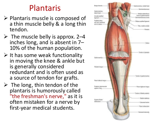
The plantaris is a small muscle that courses along the posterior aspect of the leg as part of the postero-superficial compartment of the calf. The muscle itself is quite small and is located in the superior aspect of the calf, attaching from the posterior femur and crossing the knee medially (as shown in the image below).

The plantaris muscle and tendon (source Google images)
It then forms a long tendon which courses down the back on the leg between the soleus and gastrocnemius. It continues inferiorly along the medial aspect of the soleus muscle belly, to the Achilles tendon, which it accompanies to its insertion on the medial aspect of the calcaneus. Anatomic studies have shown that the calcaneal insertion (heel bone) may also occur independently of the Achilles tendon (1). This is of interest as the plantaris tendon will often remain intact when the Achilles tendon ruptures (2). The plantaris, together with the gastrocnemius and soleus, are collectively referred to as the triceps surae muscle.
Patient History – Assessment of Plantaris pain
Although injuries of the plantaris have been a source of controversy over many years, pathology of the plantaris muscle and tendon is an important differential diagnosis for pain arising from the posterior aspect of the leg (3). To help differentiate between Achilles and plantaris tendon pain, there are a few key notes from the client’s history.
What to Look For
Pain location of plantaris tendon pain is medial, where the plantaris sits adjacent to the Achilles. Pain is typically 6-8 cm above the Achilles insertion, where the plantaris merges with the Achilles tendon and its tendon sheath. Midportion Achilles tendinopathy will be slightly lower in the body of the tendon, whilst insertional Achilles tendinopathy will be much lower at the insertion point of the Achilles on the calcaneus. Pain location can be a key driver to help identify plantaris pathology (3).
The history of Achilles pain will also be an important consideration. Plantaris pain often has a sudden onset during a running based activity. It can occur in isolation without strong pain or alongside gastrocnemius or soleus muscle strains (tears). This is common with a fast, heavy eccentric load (lowering body weight) placed across the ankle with the knee in an extended position. Often this may feel like you’ve been struck on the calf by an object such as a ball or piece of equipment. Depending on the severity, calf soreness may be experienced that may cause the athlete to stop play, or may simply be experienced throughout the remainder of the activity. This pain usually becomes more severe after resting or the next day. Accompanying the pain may be swelling that may extend down to the ankle and foot.
Plantaris pain often has a sudden onset during a running based activity #plantaris #physio #painfree #running #injury Share on X
Plantaris pain can also occur more gradually over time without this previously mentioned acute strain mechanism. The pattern of symptoms is often similar to an Achilles tendinopathy, with start up pain, then decreasing with greater duration physical activity. This is as the plantaris tendon can become tendinopathic, which results in structural changes mirroring other tendinopathies such as neovascularisation of peritendinous tissues (increased nerve fibres surrounding the tendon). This has been proposed to have links to changes and pain from the Achilles tendon, however the extent of this interaction is yet to be fully understood. The tendinopathic changes thus can be imaged using Ultrasound Tissue Characterisation (UTC) or musculo-skeletal ultrasound.
Examination Findings – Assessment of Plantaris Pain
On examination pain will be reproduced over the plantaris tendon. The tendon, which is approx 3mm in diameter, is palpated where it exits from the muscle tendon junction between the Gastrocnemius and the Achilles and followed down on the medial aspect, noting the site of maximal tenderness.
Individuals with plantaris pain, similar to Achilles tendinopathy will describe pain loading into dorsi-flexion (heel raise). Assessment of body-weight dorsi-flexion of a heel raise may be painful and more painful with a straight knee versus a bent knee. If pain is unable to be reproduced your physiotherapist may move to single leg calf raises, repeated calf raises or single leg hops, noting the location of pain when provoked.
Further assessment will also serve to rule out other potential diagnoses; such as gastrocnemius or soleus strains, Achilles tendon ruptures, referred pain or retrocalcalneal bursitis. Contributing factors will also be examined such as foot posture, footwear and dorsiflexion range-of-movement (ROM). Decreases in dorsiflexion ROM may increase pronation and subsequent compression on the medial aspect of the Achilles.
Decreases in dorsi-flexion ROM may increase pronation & subsequent compression on the medial aspect of the Achilles #physio #plantaris Share on X
Treatment of Plantaris & Achilles Tendinopathy
Surgery is the only treatment currently studied for Plantaris, although clinically, conservative management is often successful. Surgical resection of the plantaris has been show to improve tendon structure and decrease pain (5). If the injury occurs acutely alongside muscle strains typical initial management should be utilised; rest from aggravating activities, ice, calf muscle compression, elevation (if swollen) and gradual reintroduction of normal movement patterns.
Conservative Management of Plantaris Pain
For managing plantaris pain presenting like a tendinopathy, management will be similar to an Achilles insertional tendinopathy. Initially isometrics can be helpful in reducing tendon pain. Management will then progress to through range strengthening, as calf/heel raises from flat ground, not off the edge of a step. If the patient has pain into dorsiflexion, initially management will involve avoiding loading down into dorsiflexion (eccentrics). Similarly, if this occurs during running dorsiflexion may also be reduced, by changing stride length, cadence and or foot placement (see HERE for related blog posts to running technique). Gradual progression to faster, higher loads through the tendon will occur guided by your physiotherapist.
Treating plantaris tendinopathy may also incorporate changes in footwear and incorporate heel raises to help de-load compression of the tendon. Additionally manual therapy and taping may be useful for Plantaris to settle down the tendon. Click HERE for a framework on how to best select running shoes.
The plantaris is an important factor to be considered and treated to successfully manage Achilles tendon pain. If you are unsure about best diagnosis and best management please contact your local Physiotherapist to get you back on track to better and pain-free performance.

Lewis Craig (APAM)
Physiotherapist
POGO Physio
References
(1) Kirwan, P. D., French, H. P., & Duffy, T. (2015). PLANTARIS MUSCLE, ITS LOCATION AND SIZE IN THE REGION OF THE ACHILLES TENDON: AN OBSERVATIONAL CADAVERIC STUDY. Bone & Joint Journal Orthopaedic Proceedings Supplement, 97(SUPP 11), 18-18.
(2) Spina, A. A. (2007). The plantaris muscle: anatomy, injury, imaging, and treatment. The Journal of the Canadian Chiropractic Association, 51(3), 158.
(3) O’Neill, S. (2015) Physio Edge 042 Treatment of Plantaris & Achilles Tendinopathy with Seth O’Neill., D. Pope.
(4) Spang, C., Alfredson, H., Harandi, V. M., & Forsgren, S. (2014). 97 Richly Innervated Tissue In Between The Plantaris And Achilles Tendon In Achilles Tendinopathy. British Journal of Sports Medicine, 48(Suppl 2), A63-A63.
(5) Masci, L., Spang, C., van Schie, H. T., & Alfredson, H. (2015). Achilles tendinopathy—do plantaris tendon removal and Achilles tendon scraping improve tendon structure? A prospective study using ultrasound tissue characterisation. BMJ Open Sport & Exercise Medicine, 1(1), e000005.
![]()
If you are ready to say goodbye to pain and injury and hello to your pain free best performance, get started with our Discover Recover Session, click HERE to schedule.


