Insertional Achilles Tendinopathy

Insertional Achilles Tendinopathy is a painful and difficult condition that many runners and active individuals experience. Importantly insertional achilles tendinopathy (IAT) is a different pathology to other types of achilles pain and as such it may not respond to treatments given to other types of achilles presentations. It has been estimated that one third of achilles pain is IAT (1,2). Here we discuss some key features of recognising IAT and important aspects of its management.
Anatomy
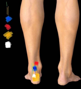
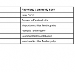
The large achilles tendon inserts onto the back of the heel bone (calcaneum). Sitting in behind the achilles at the insertion is the retrocalcaneal bursa. The achilles tendon, the fibrocartilage walls of the bursa and the cartilage surface of the heel bone form a complex interlinked attachment point (enthesis) (3). Bone spurs are a common finding in foot and achilles pain and those without symptoms. The presence of the osteophytes is not itself considered a contributing factor in IAT although an increased size may play a role in symptoms (2). Degenerative changes within the Achilles insertion with pain in this exact location is the hallmark of IAT. Tendon degeneration is marked by a loss of parallel collagen structure, loss of fiber integrity, fatty infiltration, and capillary proliferation (4).
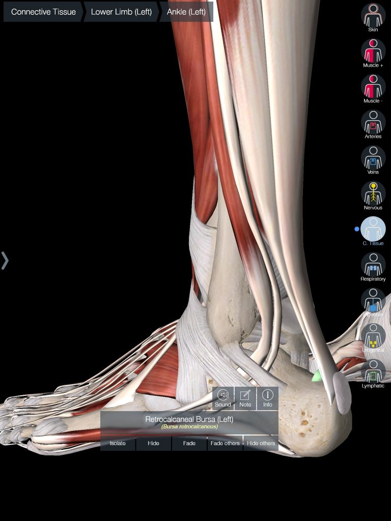
Presentation
IAT has a slightly different presentation to other forms of Achilles pain. Its location is lower; within 2cm from the insertion point on the heel and often on the outside of the achilles (2). There is often an area of redness or swelling at the pain site. Due to its location, insertional achilles tendinopathy is often aggravated by compressive loads such as stretching the calf into dorsiflexion (think knees moving past your toes). These presentations will be more likely to be aggravated from decline calf raises (heels below ground level such as off a step) (4) and will prefer to be in shoes rather than barefoot. In the early stages, pain is often reported that their symptoms occur only after strenuous activity, or at the beginning of activity then improves. As symptoms progress less strenuous activity may cause symptoms or the pain may not improve or warm up. Symptoms could even then occur without activity. Patients may even experience symptoms at rest.
Onset is typically after a period of increased compressive loads such as loading into dorsiflexion as mentioned above. This may also occur if someone has changed shoes and quickly moved from a high drop shoe (eg 8-10mm drop) to a low drop (0-2mm) drop shoe. Alternatively, it may occur slowly following a rapid increase in tensile loads such as an increase in running volume and/or intensity. Patients with IAT often report stiffness that is aggravated by prolonged rest as well as pain that is aggravated by physical activity, with activity that places more load through the achilles creating higher levels of pain. In addition, because of sensitivity over the posterior heel, many struggle with shoe wear (2).
Management
IAT management is primarily non-surgical and rehabilitated with a progressive strengthening or loading program. It involves graded adaption to compressive and tensile loading through a variety of exercise choices. A common protocol for calf loading is eccentric training where heavy resistance is applied in a ‘lowering calf raise over the edge of a step.’ A more recent study has found performing this without moving into dorsiflexion (no-step) is a promising method of reducing achilles pain (5). Although this study looked at eccentric contraction, more recently the approach for graded loading of the achilles is typically focused on factors such as maximum load, speed of contraction, and frequency of sessions, rather than mode of contraction (eccentric vs concentric) (6). This then enables exposure to graded all loads, including plyometric loads and running. Wearing shoes with a higher heel or external wedge can often be helpful for these cases as well (2).

Lewis Craig (APAM)
POGO Physiotherapist
Masters of Physiotherapy
Featured in the Top 50 Physical Therapy Blog
References:
- Karjalainen PT, Soila K, Aronen HJ, et al. MR imaging of overuse injuries of the Achilles tendon. Am J Roentgenol. 2000;175(1):251-260.
- Chimenti, R. L., Cychosz, C. C., Hall, M. M., & Phisitkul, P. (2017). Current Concepts Review Update: Insertional Achilles Tendinopathy. Foot & Ankle International, 38(10), 1160–1169. https://doi.org/10.1177/1071100717723127
- Rufai, A., Ralphs, J.R. and Benjamin, M. (1995), Structure and histopathology of the insertional region of the human achilles tendon. J. Orthop. Res., 13: 585-593. https://doi.org/10.1002/jor.1100130414
- Movin T, Gad A, Reinholt FP, Rolf C. Tendon pathology in long-standing achillodynia. Biopsy findings in 40 patients. Acta Orthop Scand. 1997;68(2):170-175
- Jonsson P, Alfredson H, Sunding K, et alNew regimen for eccentric calf-muscle training in patients with chronic insertional Achilles tendinopathy: results of a pilot studyBritish Journal of Sports Medicine 2008;42:746-749.
- Malliaras P, Barton CJ, Reeves ND, Langberg H. Achilles and patellar tendinopathy loading programmes: a systematic review comparing clinical outcomes and identifying potential mechanisms for effectiveness. Sports Med. 2013;43(4):267-286


